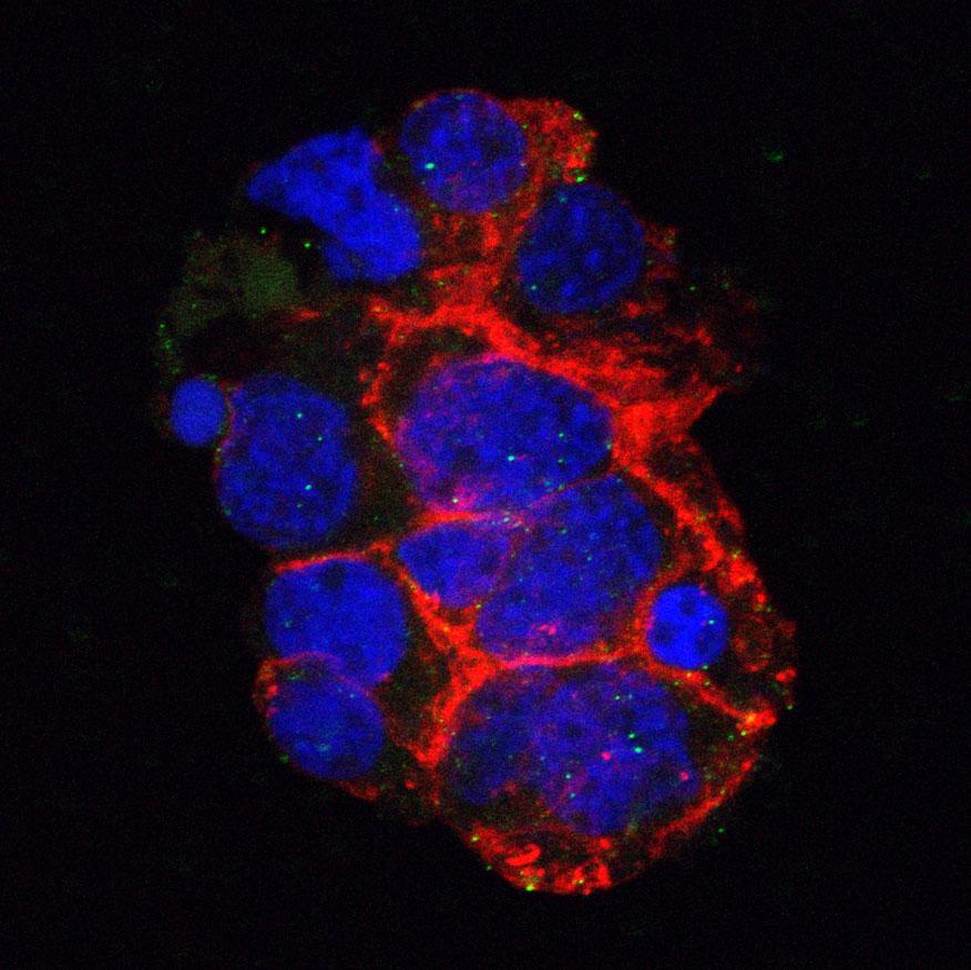
Lumpy skin disease virus (LSDV) causes lumpy skin disease (LSD) in cattle and buffalo.
Other wild ruminants have been reported to be susceptible to LSDV, such as springbok, impala, giraffe, camel, Indian gazelle, and yak.
The exact mechanisms of LSDV transmission between animals and herds are not fully understood. Strong evidence suggests that the virus is spread by biting insects such as flies (e.g. Stomoxys calcitrans and Biomyia fasciata) and mosquitoes (e.g. Culex mirificens and Aedes natrionus). While direct transmission between animals is believed to occur, its significance compared to vector transmission remains unclear.
Lumpy skin disease (LSD) was originally found in southern Africa but has spread northwards over the past decade. The disease is endemic in most African countries.
Before 2012, only sporadic outbreaks occurred in the Middle East, but since then, LSDV has spread to Lebanon, Jordan, Saudi Arabia, Iran, and Turkey. From there, it moved into Russia (Dagestan), Armenia, Greece, Bulgaria, and the Republic of North Macedonia. Though controlled in Eastern Europe through vaccination, LSDV has continued its spread across South-Eastern Asia, including India, China, Mongolia, Thailand, and Indonesia.
Please see the Defra website for advice on how to spot and report the disease.
Clinical signs
Clinical signs vary, with younger animals more severely affected.
- Nodular skin lesions (lumps) on the animal’s body, muzzle, nose, head, neck, back, legs, scrotum, perineum, udder, eyelids, tail and mouth
- Nodules can also develop internally, particularly in the respiratory and gastrointestinal tracts
- Fever
- Listlessness and reluctance to eat
- Ocular and nasal discharge
- Milk drop with weight loss
Virology
Taxonomy
Realm Varidnaviria, kingdom Bamfordvirae, phylum Nucleocytoviricota, class Pokkesviricetes, order Chitovirales, family Poxviridae, subfamily Chordopoxvirinae, genus Capripoxvirus, species Lumpy skin disease virus
Virion
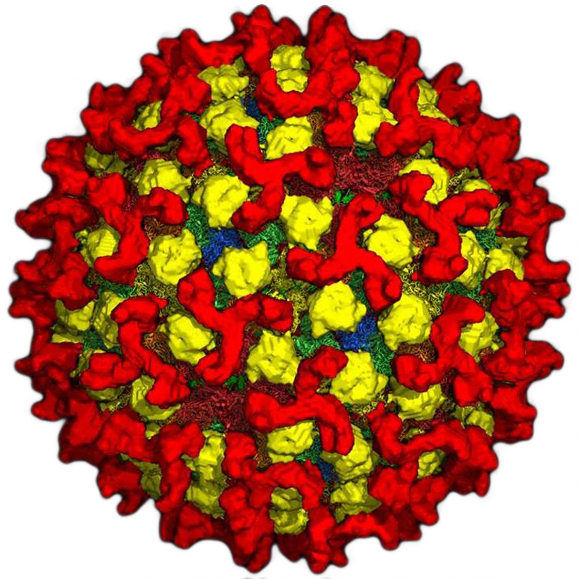
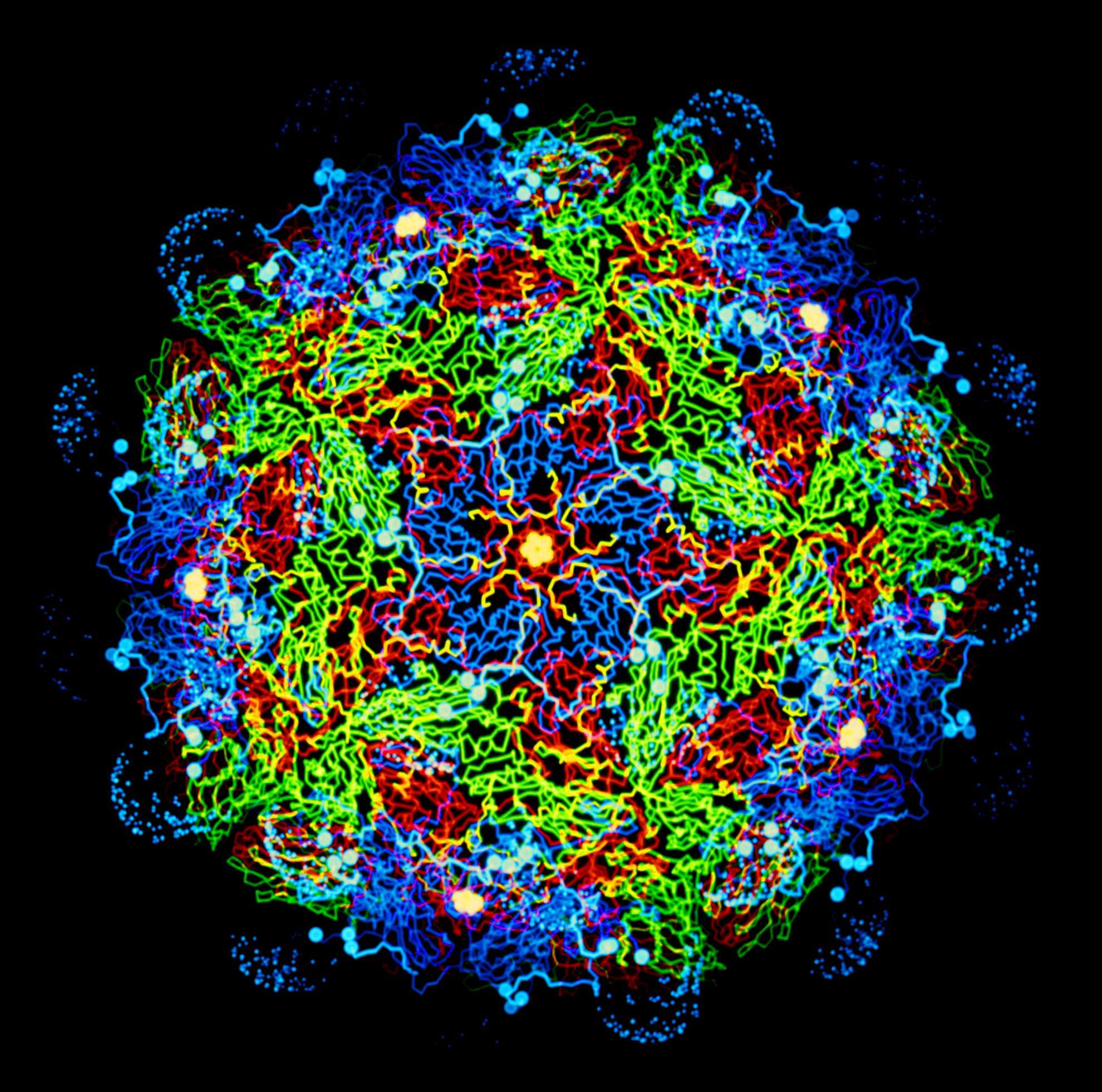
LSDV virion displays a typical poxvirus morphology: brick-shaped virions (220-450 long × 140-260 wide × 140-260 nm thick). Virions consist of a lipoprotein surface membrane enclosing a biconcave core that contains the DNA genome. Two lateral bodies are present in the concave regions between the core and the membrane.
Genome
The LSDV genome is a linear molecule of dsDNA,151,000 base pairs (bp) in length, with covalently-closed ends. The viral genome encodes 156 putative genes and is organized in a central region flanked by identical inverted terminal repeats of ~2,400 bp each.
Lifecycle
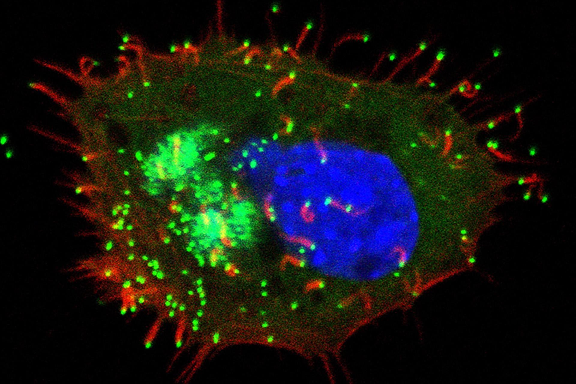
Virus entry is mediated by fusion between viral and cellular membranes at the plasma membrane or following endocytosis.
The early phase of replication includes expression of proteins needed for replication of viral DNA and modulation of the host cellular functions and antiviral defences.
DNA replication and gene expression occur in the cytoplasm in ‘virus factories’ and is mediated by virus-encoded proteins.
The late phase of replication includes the expression of structural proteins involved in the multistage assembly of new virions. Virions are released by exocytosis or after cell lysis.
Pirbright's research on lumpy skin disease
As the home of the National and World Organisation for Animal Health (WOAH) reference laboratory for LSDV, Pirbright plays a critical role in diagnostics, surveillance, and advising the UK government and WOAH.
We maintain and continuously improve molecular and serological tests to detect LSDV in infected ruminants and are currently developing new diagnostic tests to enhance detection accuracy.
Using the latest next-generation sequencing technologies, we are improving the speed and efficiency of determining the genomic sequence of LSDV isolates. This work will help us to understand how viruses evolve, spread and the origin of new incursions or outbreaks.

