Our group
Bioimaging at The Pirbright Institute brings together cutting edge instrumentation and a dynamic, experienced team to provide exciting collaboration opportunities.
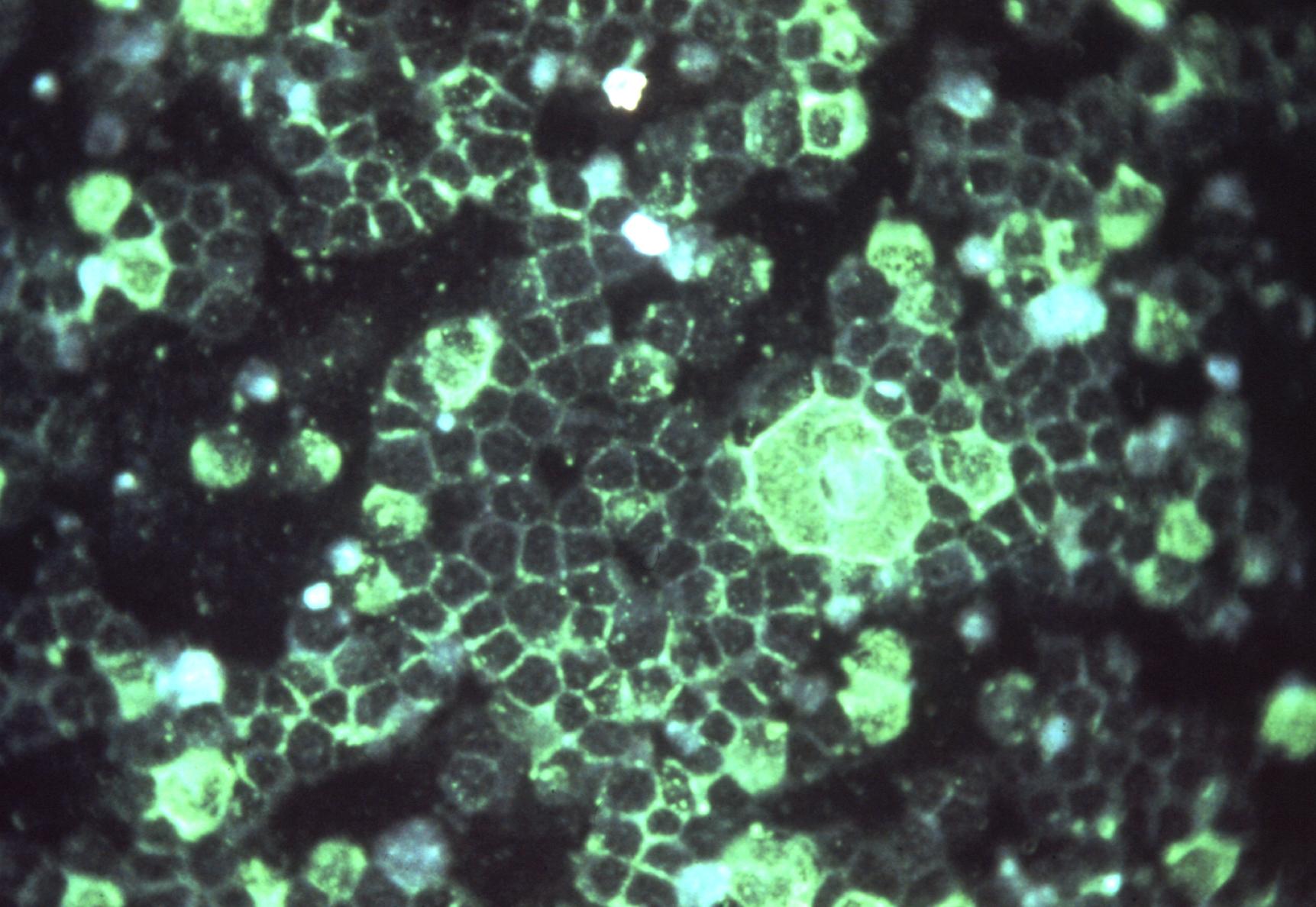
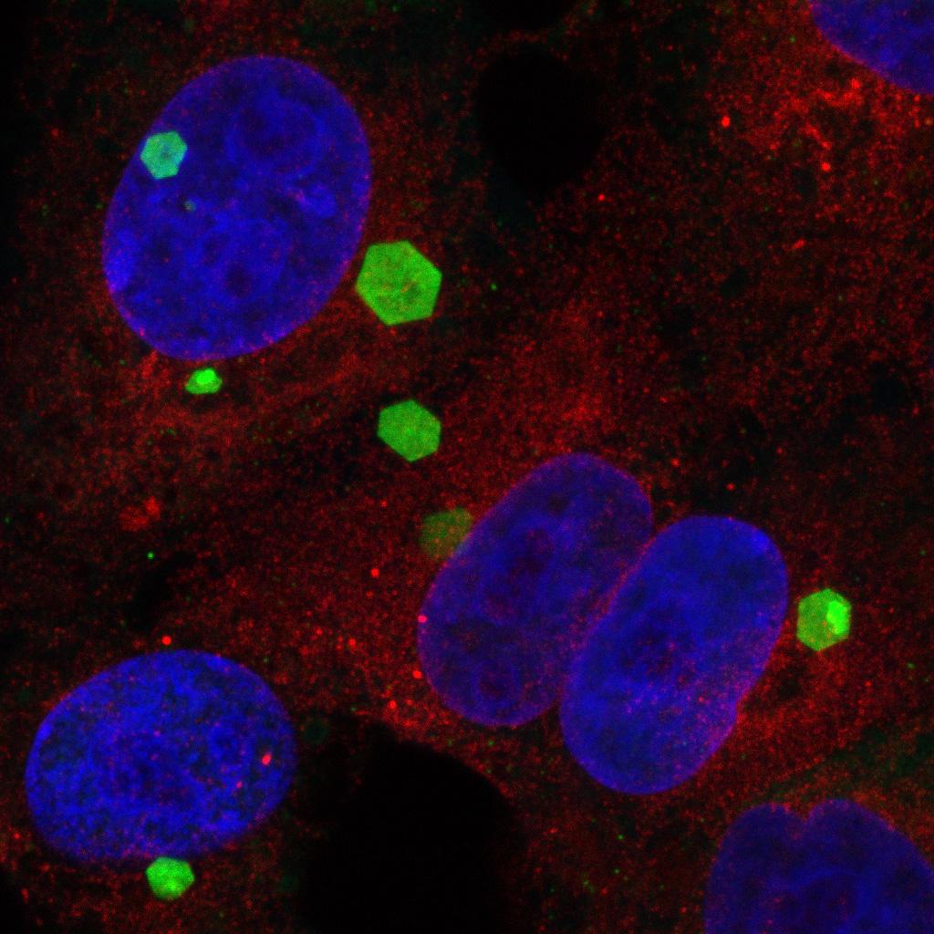
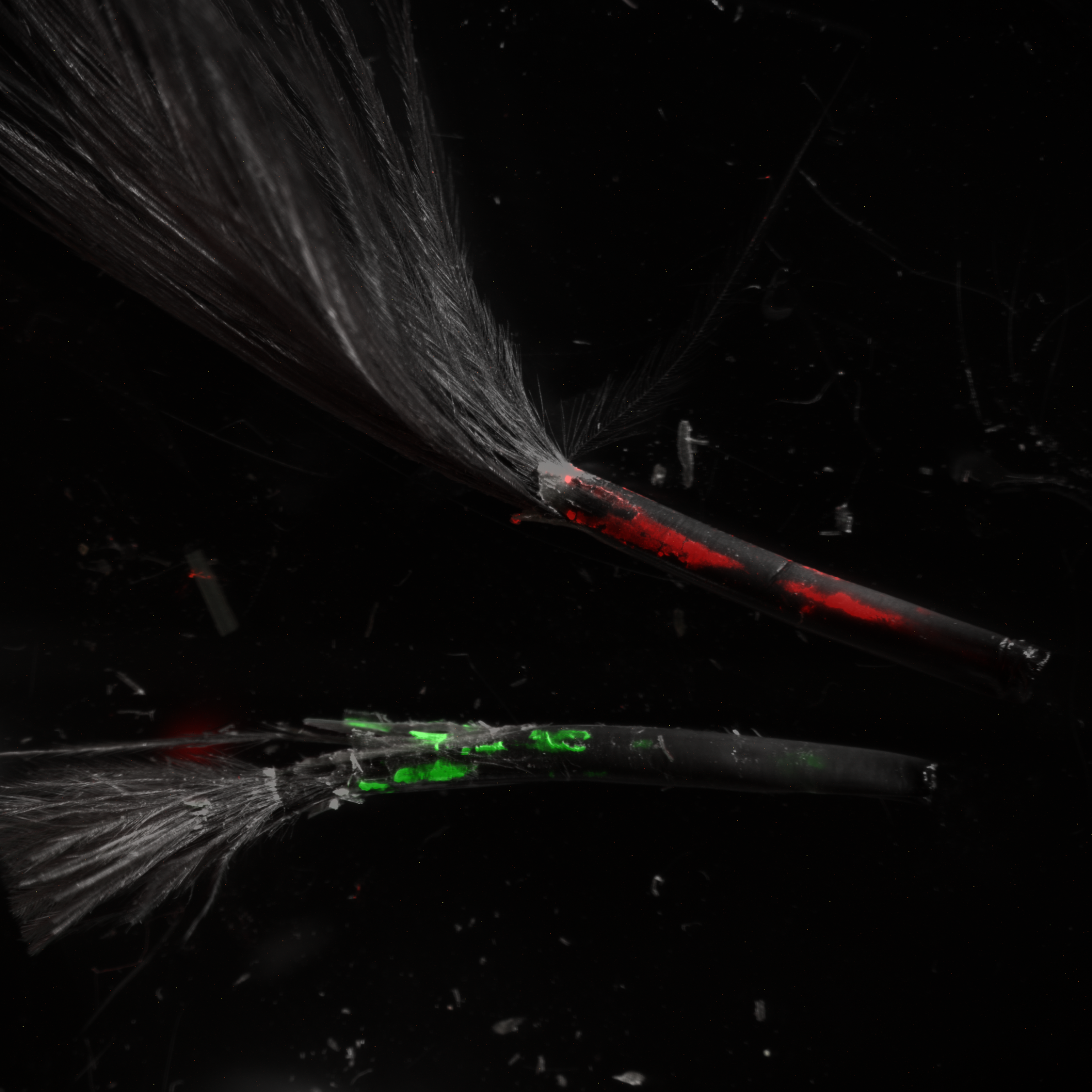
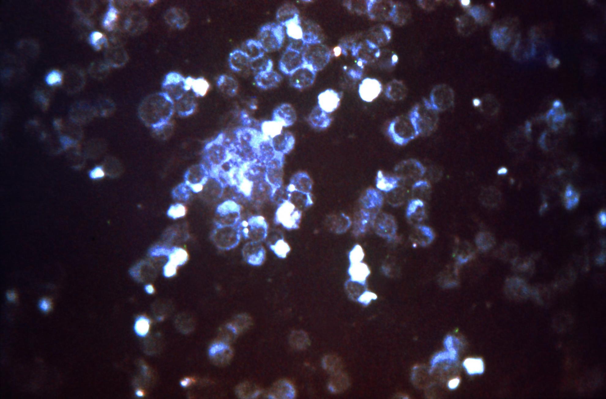
High resolution microscopes provide the opportunity for researchers to visualise viruses in situ and identify the location of molecules within cells. Bioimaging suites are located in both high and low containment environments (The Plowright Building and The Jenner Building respectively), making them accessible to all researchers. The instruments located within both Bioimaging Suites provide a unique environment for the study of animal and zoonotic pathogens and are available for use by both internal and external collaborators.
Our instrument portfolio is detailed below:
The Plowright Building
- Leica SP8 CLSM with gSTED, inverted microscope for live or fixed samples
- Leica SP8 CLSM, upright microscope for fixed samples
- ThermoScientific Talos L120C G2 transmission electron microscope for negatively stained samples and resin embedded samples
- JEOL 2100F 200kV FEG transmission electron microscope for electron tomography
- Leica EM ICE high pressure freezer and AFS2 freeze substitution unit
- Leica UC6 ultramicrotome and UC7 cryo-ultramicrotome
- Leica CM1860 UV cryostat and Leica vibrating microtome
The Jenner Building
- Leica STELLARIS 5 inverted microscope for live or fixed samples
- ThermoScientific Vitrobots for vitrification of samples
- Leica CM1860 UV cryostat and Leica vibrating microtome
The Bioimaging group consists of experienced staff members who have a broad knowledge of the techniques needed to image viruses, as well as expertise in using the instrumentation itself.
Our aims
The aims of the Bioimaging group are to promote the use of microscopy in the study of animal and zoonotic pathogens and to ensure excellence in microscopy at The Pirbright Institute. The operation of cutting edge, high resolution microscopes at high containment brings many technical, practical and strategic difficulties, but it is our aim to establish Bioimaging at The Pirbright Institute as the gold standard for containment microscopy.
Our research
Bioimaging works alongside many groups within the Institute and are able to collaborate with researchers within the UK and overseas. Collaborations have included work on foot-and-mouth disease virus, infectious bronchitis virus, African swine fever virus, rinderpest virus, bluetongue virus, Marek’s disease virus, Hendra virus (with AAHL, Geelong, Australia) and pox viruses.
Bioimaging staff members are also involved in the promotion of microscopy including teaching, training and encouraging schools to include microscopy as part of their curriculum.
Our impact
Virus infection of host cells can be broken down into several broad steps; entry, genome replication, morphogenesis and exit. Full understanding of these processes is needed before we are able to manipulate them to prevent viral spread.
Microscopes are used to localise viral proteins and identify interactions at a host cell level so we can understand how viruses use their environment to proliferate. This fundamental research can be used to develop procedures or reagents which block infection, therefore providing the basis for an anti-viral treatment or vaccine. Vaccines and anti-viral treatments for animal pathogens reduce financial losses for farmers, increase productivity and enhance the health of the animal population.